Postdoctoral Research
Oyen Lab at Wayne State University
Analysis of placenta ultrasonic images
Fetal growth restriction occurs in an estimated 5% of pregnancies and in many cases may be caused by placental issues. In this project, we attempt to improve prediction of which pregnancies will experience fetal growth restriction by analyzing the texture of the placenta in ultrasonic images, using established radiographic and machine learning methods.
Cesarean section scar computational models
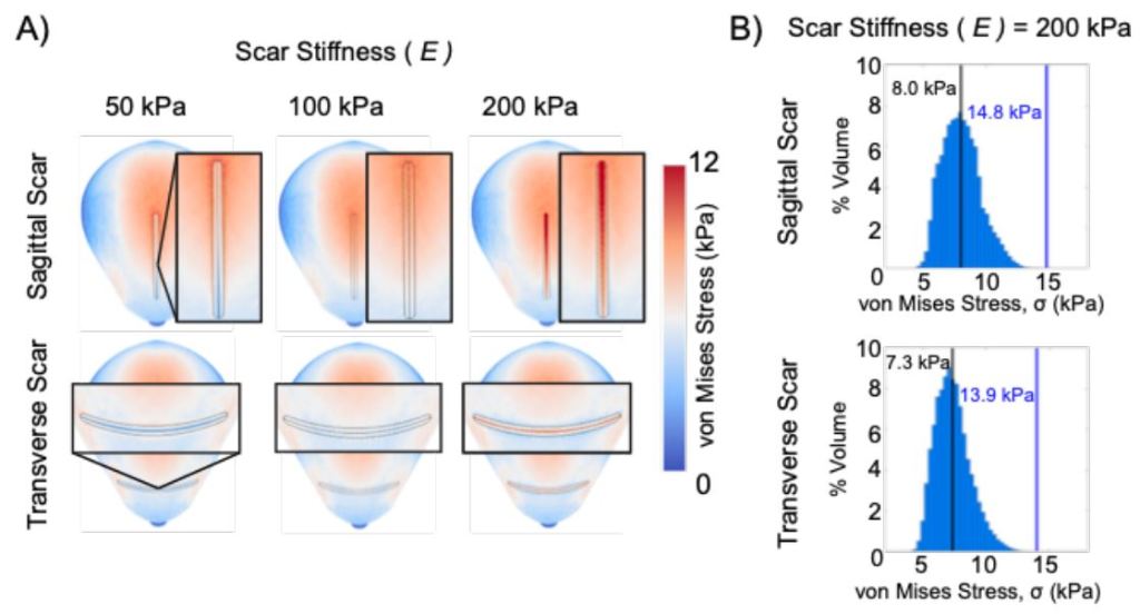
Cesarean section is a common mode of delivery, but it is known that there is increased rates of uterine rupture and preterm birth in subsequent pregnancies. Using finite element models of pregnant reproductive tissues, we parametrically investigate the effect of cesarean section scars on tissue loading.
Modeling placental abruption
Abruption of the placental is essential for a healthy delivery, where the placenta separates from the uterine wall. However, placental abruption before delivery can have devastating consequences for the mother and fetus. One of the most acute scenarios where placenta abruption can occur is in car crashes, thus in this project we aim to create a model of placental abruption to study how it can be avoided in car crashes.
Doctoral Research
Myers Soft Tissue Lab at Columbia University
Patient-specific models
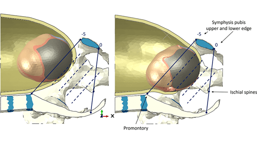
I have developed a method to model the uterus and cervix on a patient-specific basis. The models are based on measurements taking from 2D ultrasound images, and then automatically generated using a design-table driven approach in Solidworks. This method can be used to model anatomy across gestational age.
Finite Element Analysis (FEA) workflow
I worked on finite element analysis workflow that will calculate mechanical loading of reproductive tissues based on patient-specific solid models, patient-specific cervical stiffness, and material models based on existing literature. The workflow was used to investigate loading of reproductive tissues throughout gestation, and how certain mechanical tissue failure, such as cervical funneling, is initiated and propagated.
Clinical Studies
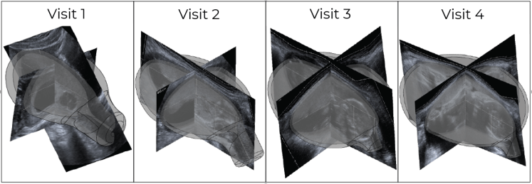
With our collaborators in Obstetrics and Gynecology at Columbia University Irving Medical Center, we are conducted a two-cohort clinical studies to asses maternal anatomy with gestational age, with low- and high-risk for preterm birth study groups. I attend patient visits to assist clinicians, sonographers, and research staff in the collection of vaginal specimens, 2D ultrasounds, and cervical stiffness measurements. These data will be used in our FEA workflow to study difference in maternal anatomy and loading for patients at low- and high-risk for preterm birth.
Collaborations
Professor Antoine Jerusalem & Alice Collier
Oxford University
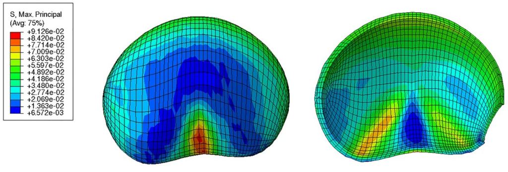
Investigating fetal head molding during vaginal birth, published in ASME Journal of Biomechanical Engineering (https://doi.org/10.1115/1.4065557).
Professor Michelle Oyen & Dr. Adrienne Scott
Washington University
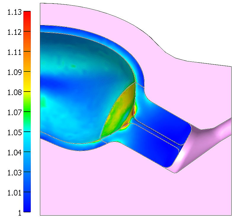
Modeling the effect of cesarean scars in the at-term uterus, published in Lecture Notes in Computational Vision and Biomechanics (https://doi.org/10.1007/978-3-031-55315-8_8).
Professor Dulce Oliveira and Daniel Fidalgo
Institute of Science and Innovation in Mechanical and Industrial Engineering
Mechanical effect of uterine contractions on reproductive tissues, published in Biomechanics and Modeling in Mechanobiology (https://doi.org/10.1007/s10237-024-01853-3).